Blog
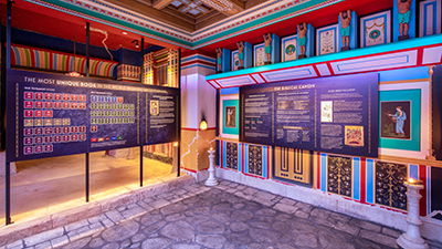
The Most Unique Book in the World
The Bible has been copied, translated, sold, and read more than any other book in history.
Learn More
The Creation Zoo’s Wallaby Walkabout Is Open!
The Creation Museum expansions are continuing with our Wallaby Walkabout, which is now open, and our Butterfly House, opening later this month.
Learn More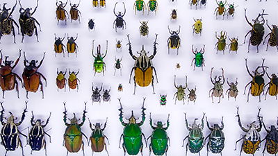
From the Beautiful to the Bizarre
God loves variety! Nowhere can we see this more clearly than among the insects.
Learn More
Rebuilding After the Flood
The global flood destroyed everything, but the descendants of Noah quickly adapted and rebuilt, showing just how intelligent and remarkable they were.
Learn More
A Family Adventure like No Other—See What’s Waiting for You!
There are so many questions about our world, and so many answers are just waiting to be discovered at the Creation Museum, using the Bible as our starting point!
Learn More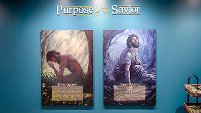
The First and Last Adam
The message of the Creation Museum can be summarized by looking at the first Adam and the last Adam.
Learn More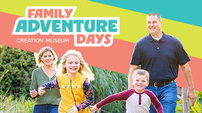
Family Adventure Days Coming to the Creation Museum
We are so excited to announce a new program at the Creation Museum: Family Adventure Days.
Learn More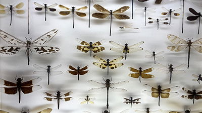
Giant Dragonflies?
The fossil record contains supersized versions of several creatures, including insects such as Meganeura, the giant dragonfly (also called a “griffinfly”).
Learn More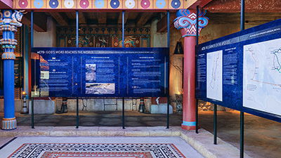
Acts: God’s Word Reaches the World
Historical and archaeological discoveries have vindicated Luke as an exceptional historian who had clearly visited the places described in the book.
Learn More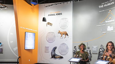
Variation Within a Kind Is Not Evolution
This variation within a kind may sound like evolution, but the two paint very different pictures.
Learn More
Made in the Image of God
After he made the animals, the Lord created the first man and woman, setting them apart by making them in his image and giving them dominion over the animals.
Learn More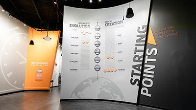
What’s the Most Reliable Dating Method?
God has always existed, knows all things, and cannot lie. Therefore, his Word is the infallible source of information regarding the age of the earth.
Learn More
How Many Animals Did Adam Name?
Adam named only “birds,” “cattle,” and “beasts of the field”—probably only animals closely associated with man and not “beasts of the earth” or “creeping things.”
Learn More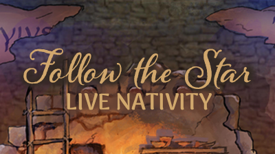
Follow the Star and Reflect on the True Meaning of Christmas
While you are here for ChristmasTown, step back in time as you view a realistic recreation of the events surrounding the night of our Savior’s birth.
Learn More
The Plants of the Bible Conservatory Is Now Open!
We’re excited to announce that our Plants of the Bible Conservatory opened to help kick off this year’s ChristmasTown at the Creation Museum on Friday.
Learn More
Double Your Impact and Help Us Upgrade the Creation Museum!
What key ministry opportunities are we looking to fund with this year’s dollar-for-dollar matching gift?
Learn More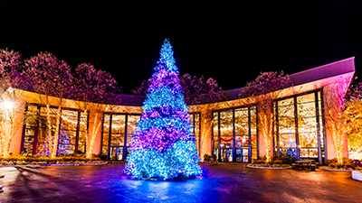
What’s New for This Year’s ChristmasTown at the Creation Museum
Beginning the day after Thanksgiving, ChristmasTown will run from 5:00 p.m. to 8:30 p.m., November 29, 2024–January 4, 2025, Tuesday through Saturday on select days.
Learn More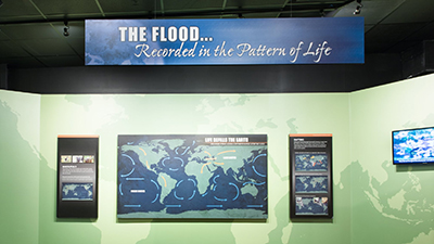
Post-flood Spread of Animal Populations
How did animals manage to quickly fill even the far reaches of the earth after the global flood of Noah’s day?
Learn More
Confusion at Babel
Babel, as the city became known, marks the historical splitting of languages and explains the language families we see evidence of in the world today.
Learn More
Global Catastrophe
The Lord was grieved at this great wickedness, and he determined to send judgment—a catastrophic global flood. But even in judgment, God chose to extend mercy.
Learn More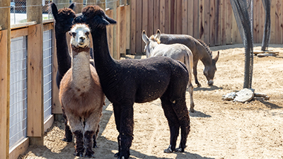
The Creation Zoo Is Open!
After nine months of closure, the day has finally arrived: the Creation Museum’s zoo, renamed the Creation Zoo, has reopened to guests!
Learn More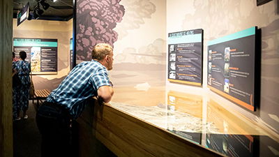
Global or Local?
Skeptics often claim the Genesis flood was merely local and not worldwide.
Learn More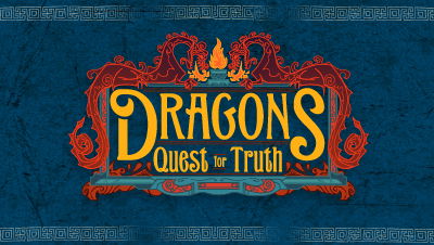
New 4D Movie Now Showing in the Special Effects Theater
A stunning new movie recently began showing in our 4D Special Effects Theater, Dragons: Quest for Truth.
Learn More
New at the Zoo!
In addition to old favorites, a number of new critters are set to call the new Eden Zoo home. Meet some of the new additions!
Learn MorePrepare to Believe
Creation Museum Location
place
2800 Bullittsburg Church Rd.
Petersburg, KY 41080 (see directions)
Seven miles west of the Cincinnati Airport
An attraction of Answers in Genesis
2025 Answers in Genesis. All rights reserved. | Privacy Policy | Content Policy | Attraction Rules